Tuberculoscope |
| A low cost fluorescence imaging device for rapid diagnosis of tuberculosis (TB) |
Overview
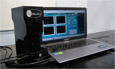
The developed device is an easy-to-use, compact and portable fluorescence imaging instrument intended for point-of-care sputum microscopy for rapid detection of Mycobacterium tuberculus (the bacteria responsible for TB disease). It acquires fluorescence images of Mycobacterium tuberculi bacteria (Mtb) from a patient’s sputum smeared on a microscope glass slide following its staining with an appropriate fluorescent dye (Auramine O). The fluorescence detection of Mycobacterium tuberculosis bacteria (Mtb) is based on the fact that the rod-shaped Mtb binds with Auramine O dye to give green fluorescence when excited with blue light.
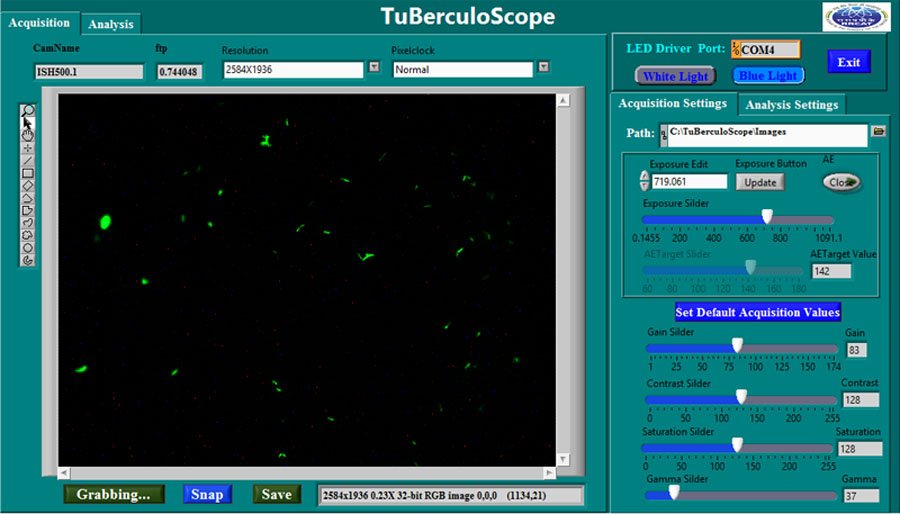
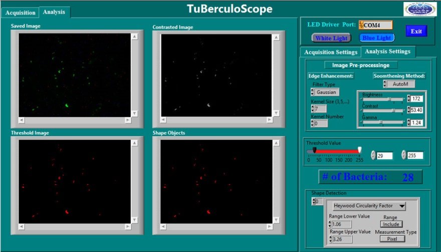
A Graphic User Interface (GUI) software enables hardware control of the device and automated counting of the Mtb bacteria in the field of view of the microscope objective. The important advantage of the device is its significantly low cost (~ 1 L) as compared to the fluorescence microscope (costing over 50 L) routinely used for TB diagnosis. This in combination with other advantages like the compactness, portability and capability of automated counting of the number of bacteria in the field of view would allow its use in a limited resource setting, particularly in remote areas thereby enabling immediate start of treatment and reduction of patient dropouts.

Quick View Leaflet
Tuberculosis (TB) is a major health concern in India. While the majority of TB cases may be treated successfully with the appropriate course of antibiotics, diagnosis remains a major obstacle to TB eradication. One of the recommended methods by WHO for the initial diagnosis of TB is based on fluorescence imaging of the rod shaped Mycobacterium tuberculus (the bacteria responsible for TB disease) from Auramine O stained sputum smear of a patient. However, the utility of this method largely remains inaccessible in rural and developing areas due to prohibitive cost of the equipment and its maintenance apart from the cost of trained technicians and laboratory infrastructure.
The developed device is an easy-to-use, compact and portable fluorescence imaging instrument intended for point-of-care sputum microscopy for detection of Mycobacterium tuberculi bacteria (Mtb). A Graphic User Interface software enables automated counting of the Mtb bacteria in the field of view of the microscope objective. The important advantage of the device is its significantly low cost (~ 1 L) as compared to the fluorescence microscope (costing over 50 L) routinely used for TB diagnosis. This along with the ease of its use would provide the advantage of putting the device in a limited resource setting in remote areas allowing rapid diagnosis followed by immediate treatment which otherwise is a time consuming procedure.
FACILITY AND MANPOWER REQUIREMENT
- Standard opto-mechanical and electronics laboratory/ workshop having assembling facilities.
One qualified Physics Graduate/ postgraduate or an opto-mechanical engineer with sound knowledge of optics, a technical assistant with working knowledge of electronics and 1-2 years of experience in assembling optical instruments and preferably a pathology lab technician having experience in working on sputum smear microscopy.
Detail Technical Brochure
Light microscopy is an essential tool for initial diagnosis of tuberculosis (TB). Unfortunately, much of the power of light microscopy, especially fluorescence imaging which is one of the recommended methods by WHO for diagnosis of TB, remains inaccessible in rural and developing areas due to prohibitive equipment and training costs. The Laser Biomedical Applications Section (LBAS) at Raja Ramanna Centre for Advanced Technology (RRCAT), Indore, a unit of department of Atomic Energy has developed an easy-to-use, compact and portable optical device - a "TuBerculoScope" for point-of-care fluorescence microscopy of the sputum smears for rapid detection of tuberculosis (TB) - suitable for resource-limited rural health services. The developed device displays fluorescence images of Mycobacterium tuberculi (the bacteria responsible for TB disease) from a patient’s sputum smeared on a microscope glass slide following its staining with an appropriate fluorescent dye (Auramine O) and illumination with blue light.
The illumination light is focused onto the dye-stained sputum-smear on the glass slide through a microscope objective and the backscattered fluorescence signal from the bacteria is collected by the same lens and detected by a CCD camera. The fluorescence detection of Mtb is based on the fact that the rod-shaped bacteria bind with Auramine O dye to give green fluorescence when excited with blue light. The resolution of the system is high enough to allow easy identification of individual TB bacteria in the sputum smear being interrogated.
A Graphic User Interface (GUI) software, developed in-house, enables hardware control of the device and automated counting of the Mtb bacteria in the field of view of the microscope objective. The important advantage of the device is its significantly low cost (~ 1.5 L) as compared to the fluorescence microscope (costing over 50 L) routinely used for TB diagnosis in clinical pathology labs. This in combination with other advantages like the compactness, portability and capability of automated counting of the number of bacteria in the field of view would allow its use in a limited resource setting, particularly in remote areas thereby enabling immediate start of treatment and reduction in patient dropouts.
FEATURES
- Low cost: Tuberculoscope (~Rs. 1.5 Lakh) vs Fluorescence Microscope (~Rs. 50 lakhs)
- Based on the principle of fluorescence microscopy
- Automated counting of number of bacteria in the field of view
- The instrument runs on a laptop battery, no extra power source is required
- Ease of use in remote areas
- Compact and portable, it can be taken anywhere.
- On-spot screening would allow immediate start of treatment thereby limiting patient drop out.
APPLICATIONS
- Rapid detection of tuberculosis (TB) through point-of-care fluorescence microscopy of sputum smears of patients suspected of having infection by Micobactrium tuberculi bacteria responsible for TB
SPECIFICATIONS
| Based on the principle of fluorescence microscopy of Auramine O stained sputum smear | |
| Mechanical Dimensions | 26.3 cm X 13.5cm X 10.5cm |
| Weight | 2.6 Kg |
| Power Input | Draws power from USB port of any computer/laptop (requires two USB ports). |
| Temperature stability | ± 1.5 °C |
INFRASTRUCTURE
- Standard opto-mechanical/ electronic laboratory with assembling facilities.
TEST EQUIPMENTS
- Test and measuring facility for LEDs like spectrometer, Digital Voltmeter, DC power supply for testing, PC with windows xx OS.
OTHER FACILITIES
- Standard opto-mechanical/ electronic laboratory having assembling facilities of components like objective lens, sample stage, micrometer screws
POWER
- As required by interfacing computer/laptop
SKILLED MANPOWER
- One qualified Physics Graduate/ postgraduate or an opto-mechanical engineer with sound knowledge of optics, a technical assistant with working knowledge of electronics and 1-2 years of experience in assembling optical instruments and preferably a pathology lab technician having experience in working on sputum smear microscopy.


