Vision Enhancement Module (VEM) |
| A low cost fluorescence imaging tool for in-vivo screening/diagnosis of malignant and potentially malignant lesions of human oral cavity |
Overview
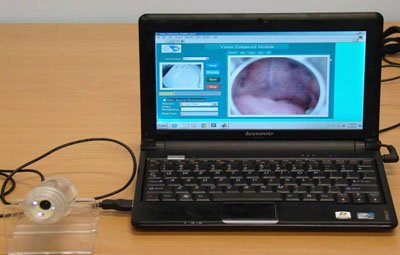
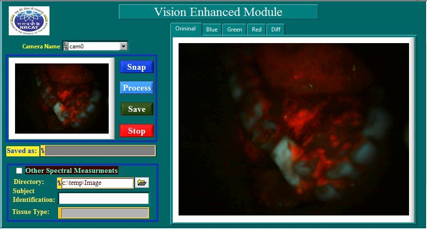
The developed device, Vision Enhancement Module (VEM), is a hand-held, USB powered fluorescence imaging tool intended for in-vivo detection of malignant and potentially malignant lesions of human oral cavity. It is a low-cost, health-care instrument capable of acquiring fluorescence images from wide area (≥1 cm2) of tissue surface. VEM uses multiple light emitting diodes (LEDs) for inducing fluorescence in native tissues. The light from the LEDs is shone onto the tissue surface to be probed and the fluorescence emitted from the tissue is detected by a CCD camera to generate two dimensional fluorescence spectral images. A Graphic User Interface (GUI) software developed and integrated with this imaging device enables automated acquisition and processing of tissue fluorescence images for highlighting the difference in the fluorescence characteristics of the different tissue types.

Quick View Leaflet
Patients diagnosed with oral cancer at an early stage have a substantially greater chance of successful treatment and less treatment - associated morbidity than those diagnosed at a late stage. Currently, the most widely used screening test for oral cancer is visual inspection of the oral cavity. However, often it turns out to be difficult to satisfactorily detect changes in oral mucosa, associated with early cancers or pre-cancerous alterations that generally precede invasive cancers, using conventional oral examination under white light illumination.
This Vision Enhancement Module (VEM), is a handy fluorescence imaging tool for real-time, non-contact and in-situ imaging of fluorescence from human oral cavity intended for improved visual assessment of the oral cavity. Using this instrument, regions of oral lesions can be better identified against the healthy oral tissues based on their natural characteristics in response to light. The method involves shining light from the LEDs of the VEM onto the tissues in the oral cavity for inducing fluorescence in the native oral tissues. The fluorescence emitted from the oral cavity, different for normal and abnormal oral tissues, is detected by a CCD camera to generate two dimensional fluorescence spectral images. A Graphic User Interface (GUI) software developed and integrated with this imaging tool enables automated acquisition and processing of tissue fluorescence images for highlighting the difference in the fluorescence characteristics of the different oral tissue types.
SPECIFICATIONS
- Mechanical Dimensions : Length 120mm, diameter 40mm
- Weight : 140 gm
- Power Input : Draws power from USB port of peripheral computer/laptop
APPLICATIONS
The developed device is meant for use in a clinical situation to better identify oral lesions as well as to improve visual assessment of the oral lesions by experienced doctors.
Detail Technical Brochure
The Laser Biomedical Applications Section at Raja Ramanna Centre for Advanced Technology, Indore, a unit of department of Atomic Energy has developed a hand-held optical tool, Vision Enhancement Module (VEM), for real-time, non-contact and in-situ imaging of fluorescence from human oral cavity. The device is capable of acquiring fluorescence images from wide area (≥1 cm2) of tissue surface. The light from a circular array of LEDs is shone onto the tissue surface to be probed and the fluorescence emitted from the tissue is detected by a CCD camera to generate two dimensional fluorescence images. A Graphic User Interface (GUI) software developed and integrated with this imaging device enables automated acquisition and processing of tissue fluorescence images for highlighting the difference in the fluorescence characteristics of the different tissue types. Since, the intensities of light emitted from healthy and abnormal tissues are found to be different, this allows easy identification of oral lesions. The developed device will help better identify oral lesions as well as improve visual assessment of oral lesions by experienced doctors.
FEATURES
- Based on the principle of fluorescence imaging from native tissue
- Based on the principle of fluorescence imaging from native tissue
- A low-cost, hand-held device
- Enables better identification of the early malignant and potentially-malignant oral cavity lesions not otherwise visible under white light illumination
- Smart phone compatible
- Runs on power drawn from the battery of the peripheral device( computer, laptop, or mobile), no extra power source required
APPLICATIONS
The developed device is meant for use in a clinical situation to better identify oral lesions as well as to improve visual assessment of the oral lesions by experienced doctors.
SPECIFICATIONS
| Sr. No. | Specification | Value |
| 1 | Mechanical Dimensions | Length 120mm, diameter 40mm |
| 2 | Weight | 140gm |
| 3 | Power Input | Draws power from USB port of peripheral computer/laptop |
INFRASTRUCTURE
- Standard opto-mechanical/electronic laboratory having assembling facilities
TEST EQUIPMENTS
- Test and measuring facility for electronic systems like Digital Voltmeter, DC power supply for testing, PC with windows xx OS.
SKILLED MANPOWER
- One qualified Physics Graduate/ postgraduate or an opto-mechanical engineer with sound knowledge of optics, a technical assistant with 1-2 years of experience in assembling optical instruments
POWER
- As required by interfacing tablet computer
 |
 |
 |
|
