OncoDiagnoScope |
| Tablet computer based, user-friendly portable optical spectroscopic device for screening/ diagnosis of oral cavity cancer |
Overview
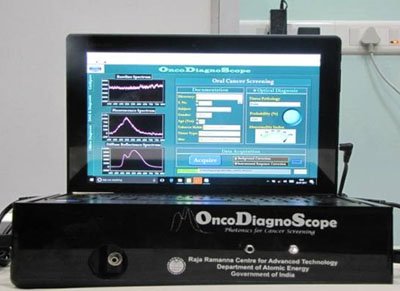
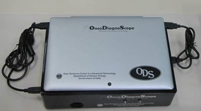
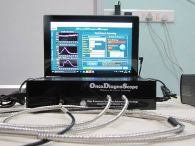
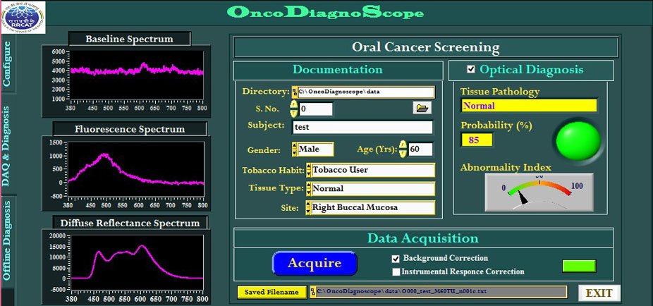
OncoDiagnoScope is a tablet computer based, compact and portable health-care instrument for real-time screening/ diagnosis of oral cavity cancer. A touch-screen enabled graphic user interface (GUI) software provides the necessary interface for the hardware control of the instrument and automation of data acquisition and analyses. During carcinogenesis, the optical properties of oral mucosa are altered. These optical changes can be measured with this USB powered machine using a pencil-sized stainless steel fiber optic probe. The fiber optic probe is brought in contact with the suspected tissue of the oral cavity of a patient and light is shone upon it. The light coming out of the tissue is captured and fed to the tablet computer where it is analyzed by a smart algorithm which can instantly determine whether the tissue is cancerous or not. The device has been validated on patients with oral neoplasia in various hospitals and cancer screening camps and found to detect cancer with an accuracy of over 90%.

Quick View Leaflet
Oral cancer is one of the most common cancers in India and its incidence is on a rise due to consumption of tobacco and pan masala. Presently, the only definitive method for determining oral cancer is through histopathological evaluation of the biopsied tissue from the suspected site. Biopsy is invasive, subject to random sampling errors and is, therefore, not an ideal screening tool. Optical spectroscopy has been suggested and validated as a powerful alternate tool for non-invasive screening of oral cancers.
The developed device is a compact and portable, optical spectroscopy based, health-care instrument intended for real-time diagnosis/screening of oral cavity cancer. With the assistance of this instrument, doctors will be able to non-invasively detect, in real-time, whether any pre-cancerous or cancerous lesion is present in the oral cavity of the subject he is examining. It uses two types of light (fluorescence and reflectance) returned from the tissue to determine its' tissue type without disturbing or destroying the tissue. The device has been validated on patients with oral neoplasia in various hospitals and cancer screening camps and found to detect cancer with an accuracy of over 90%.
FEATURES
- Based on the principle of optical (fluorescence and diffuse reflectance) spectroscopy.
- Compact and portable.
- Equipped with touch-screen enabled GUI software for ease of operation, data documentation and analyses.
- Provides instant diagnostic feed-back about the interrogated tissue sites through display of colored flashes.
- The instrument runs on peripheral tablet battery, no extra power source required.
APPLICATIONS
- The developed device equipped with user friendly GUI software is intended for use as a standalone tool for screening population at risk of having oral cancer.
- With the assistance of this new device, it is possible to non-invasively detect in near-real time whether any pre-cancerous or cancerous lesion is present in the oral cavity of a subject. The entire investigation procedure per subject using this device is less than 15 minutes as compared to several hours (~48 hrs) required by the conventional procedure of biopsy followed by histopathology.
Detail Technical Brochure
The Laser Biomedical Applications Section at Raja Ramanna Centre for Advanced Technology, Indore, a unit of department of Atomic Energy has developed a tablet computer based, compact and portable health-care instrument, called OncoDiagnoScope, for in-vivo screening/ diagnosis of oral cavity cancer. The system consists of light emitting diodes (LEDs) and a chip-based miniaturized fiber-optic spectrometer all accommodated in a rectangular acrylic house and SMA connected to a custom-designed, pencil-sized stainless steel fiber optic probe having three legs each comprising a fused silica fiber. A miniaturized electronic data acquisition card mounted on the base of the house powers up the LEDs and is interfaced with the tablet computer fitted on the top of the acrylic house through its USB ports. A touch-screen enabled graphic user interface (GUI) software provides the necessary interface for the hardware control of the whole system and automation of data acquisition and data analysis. To measure the optical signals the fiber optic probe is brought in contact with the suspected tissue of the oral cavity of a patient. The light coming out of the tissue is captured and fed to the tablet computer where it is analyzed by a smart diagnostic algorithm which can instantly determine whether the tissue is cancerous or not. The algorithm also generates the posterior probability of diagnosis of the interrogated tissue site. Based on the probability output that the algorithm provides, red, green or orange flash is displayed with red and green implying confirmed (probability >cutoff decided by the physician) abnormal and normal respectively, while the orange indicating doubtful. The entire investigation procedure per subject using this device is less than 15 minutes as compared to several hours required by the conventional procedure of biopsy followed by histopthaology. Using this device, oral lesions can be accurately separated in a non-invasive manner from healthy oral tissues based on their natural characteristics in response to light. The device has been validated on patients with oral neoplasia in various hospitals and cancer screening camps and found to detect cancer with an accuracy of over 90%.
FEATURES
- Based on the principle of optical (fluorescence and diffuse reflectance) spectroscopy.
- Compact and portable
- Equipped with touch-screen enabled GUI software for ease of operation, data documentation and analyses.
- Provides instant diagnostic feed-back about the interrogated tissue sites through display of colored flashes.
- Provision of offline review of the spectra already recorded from different tissue sites to verify their spectroscopic impression against the clinical impression and generate a site-wise diagnostic report of the patients investigated.
- The instrument runs on peripheral tablet battery, no extra power source required.
APPLICATIONS
- The developed device equipped with user friendly GUI software is intended for use as a standalone tool for screening population at risk of having oral cancer.
- With the assistance of this new device, it is possible to non-invasively detect in near-real time whether any pre-cancerous or cancerous lesion is present in the oral cavity of a subject. The entire investigation procedure per subject using this device is less than 15 minutes as compared to several hours (~48 hrs) required by the conventional procedure of biopsy followed by histopthaology.
SPECIFICATIONS OF THE SYSTEM
| Sr. No. | Specification | Value |
|---|---|---|
| Main Unit | ||
| 1 | Mechanical Dimension | 16 cm X 12cm X 8.5cm |
| 2 | Weight | 2.5 Kg |
| 3 | Power Input | Device draws power from USB |
| Fiber Optic probe | ||
| 4 | Fiber configuration | Permanently-aligned combination of three (two excitation and one collection) fused-silica single optical fibers (600 core diameter, N.A. 0.22). The fibers running through three different legs will meet at a junction 300mm away from the distal ends of the fiber and will run through a single leg till the probe head |
| 5 | Wavelength range | 300-1000 nm |
| 6 | Probe Connectors | Fiber-optic cylindrical probe head (diameter 5 ± 0.5 mm, length 200 ± 5 mm) enclosed in stainless steel body fitted with a quartz flat of thickness 2 ± 0.2 mm at the tip |
| 7 | Probe end | Standard SMA (premium-grade SMA905 connectors) termination at both the excitation and the collection fiber(s) ends. |
| 8 | Tubing | Properly jacketed to resist damage via physical shock |
| 9 | Cable length | 2 meter |
INFRASTRUCTURE
- Standard opto-mechanical laboratory with assembling facilities.
TEST EQUIPMENTS
- Test and measuring facility for electronic systems like Digital Voltmeter, DC power supply for testing, PC with windows xx OS.
SKILLED MANPOWER
- One qualified Physics Graduate/ postgraduate or an opto-mechanical engineer with sound knowledge of optics, a technical assistant with 1-2 years of experience in assembling optical instruments.
POWER
- As required by interfacing tablet computer.
 |
 |
|
|
|
 |
|

